Localization-based super-resolution methods rely on physiological flow to relocate or replenish sparsely distributed microbubbles inside. This results in long acquisition times in the order of seconds to generate a single super-resolved frame (excluding the data processing time) due to the slow flow in microvasculature.
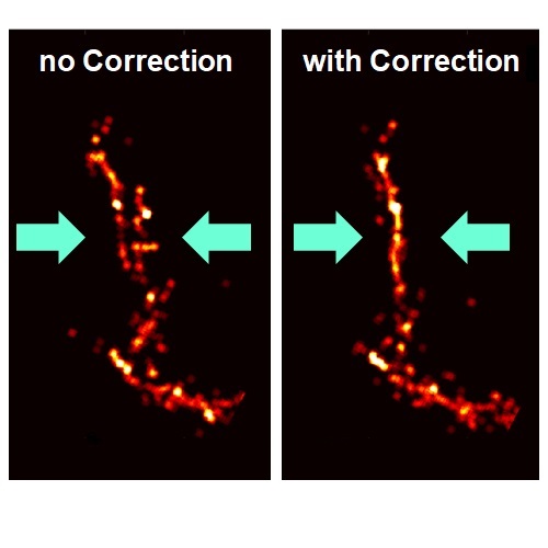
A clinical method will require a handheld ultrasound scan. Through multiple frames acquired over a duration of seconds, handheld probe movement, respiration, and cardiac motion will significantly degrade the image quality and resolution. Using retrospective data, it was possible to implement a [2D motion correction algorithm for super-resolution ultrasound imaging] (https://ieeexplore.ieee.org/document/8334280). This will form the basis of a 3D motion correction algorithm to be applied on newly acquired images. This project requires finding most suitable motion estimation and correction method for ultrasound imaging particularly for 3D super-resolution imaging.