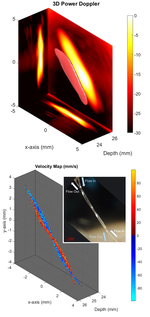
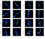
Super-resolution ultrasound is a microvascular imaging modality that can enable real-time microscopic imaging in deep tissue. Super-resolution has already been successfully demonstrated pre-clinically and clinically by Dr Harput’s previous research group. Although it is a revolutionary imaging modality, there are significant challenges for the clinical translation, such as huge amount of data and the corresponding computing power required to process such data.
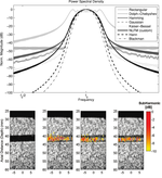
Implementing a fast in vivo imaging is essential for clinical translation. Currently, super-resolution data processing and image formation take >6 hours, which must be reduced to a clinically acceptable timescale. The existing super-localization and filtering methods must be replaced with algorithms that have lower computational complexity. Also these algorithms should be implemented on a GPU while using computer learning-based methods for further acceleration.


